
In our Harmony in Orthodontics article at right, Dr. Razdolsky talked of how 3D cone beam improves diagnostic and treatment in orthodontics, but did you know this type of dental imaging can also save lives? That’s right… 3D CBCT imaging when combined with computer aided diagnostics or (CAD) systems-based technologies can not only help dental radiologists see and diagnose various oral pathogens from dental caries (tooth decay) to cancer, they can also help prevent a stroke or hip fracture. Through expanding the scope of panoramic exams, 3D CBCT becomes a relevant screening method for osteoporosis, other cancers and even Carotid artery calcification!
Dr. Razdolsky uses CBCT in orthodontics for evaluation of bone structure and tooth orientation, as well as surgical planning for impacted teeth and even diagnosing temporomandibular joint disorder (TMJ). But it is in locating new regions of interest (ROI), or spotting suspicious signs and classifying the findings, especially when we widen the image scope to include the head and neck, we can see even greater benefit. It is then that we can allow for detection of more pathologies in one image, thereby increasing the advantage for dentists and patients.
“It’s true that we can see more than ever before, especially someone who is trained to look for such findings,” said Dr. Razdolsky. “For example, maxillary sinusitis is often caused by a dental pathology when effected roots are too close to the maxillary sinus. An experienced dentist or orthodontist will look for this and can adjust treatment or call in other specialists as needed.”
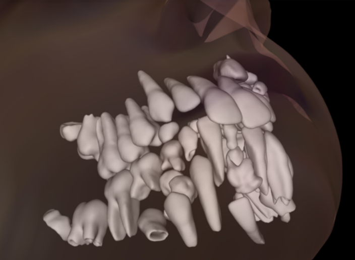
In one case, Dr. Razdolsky discovered a patient with multiple compound odontomas, or tumors. Although composed of normal dental tissue, these developmental anomalies grow in an irregular way and other than causing orthodontic pathologies, have also been linked to Gardner’s syndrome. Testing to rule out this syndrome is highly recommended as these patients can develop colorectal polyps and have an increased risk of developing colon cancer.
Another medical condition which can be evident to the trained eye is osteoporosis, a medical condition characterized by the loss of bone mineral density which increases bone fragility. This is particularly interesting as the tests to diagnose osteoporosis require special equipment and that can limit the availability of tests for the majority of patients. Since it has been proven that measuring bone mineral density in the area of the mandible can also provide accurate results in detection, panoramic dental x-rays are a much more reasonable options for screening.
“While measuring bone mineral density may not be easy for dental radiologists, the automation of CAD systems does help a lot,” said Dr. Razdolsky. “This puts dentists in a unique position to help older patients through screening and sharing that information back to the primary care physician for follow up.”
Dr. Razdolsky said the same CAD systems can be programmed to look for carotid artery calcification screening which can be spotted in dental panoramic x-ray images.
“Carotid artery calcification is a symptomless disease with potentially devastating consequences,” said Dr. Razdolsky. “A simple modification to an otherwise routine scan could save a life and just another example of how oral health can be tied to overall systemic health. As physicians, we are all in this together.”