
Dr. Razdolsky has always been an avid science and technology buff. And why not? Technology not only continues to improve the ways in which we live, at Forever Smiles it impacts how we can best serve patients through orthodontic treatment. Once such example is 3D Cone Beam technology. Since the introduction of 3D x-rays in orthodontics, Dr. Razdolsky and the team can see in greater detail structures of the mouth, jaw and developing teeth with clear and realistic imagery. This allows them to identify and predict potential dental and orthodontic conditions before they become problematic. This means treatment is proactive rather than reactive. That’s pretty cool.
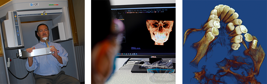
But let’s look deeper. Unlike 2D x-rays which are taken as single images from varying locations throughout the mouth, 3D imaging is considerably more convenient to a patient’s in-office visit as the images are captured in a single sweep of the iCat machine. During the scan, a module called a gantry rotates around the head, producing a buzzer sound while simultaneously capturing and combining a series of flat images from radiation beams which are stacked, combined, and digitally output to a computer to produce a 3D virtual impression of your teeth and jaw. From there, Dr. Razdolsky can develop 3D CAD-CAM models where he can rotate, shift and even digitally realign your teeth and bone structure to determine just how your teeth will look once you have successfully completed treatment. Now that’s really cool!
Wait…what? Did we say radiation? Is 3D safe?
Yes, 3D cone beam technology has been used in dentistry and orthodontics for several years now and is very safe. While 3D images do make use of many flat x-ray captures, which means more radiation is used to create the final image, the Forever Smiles team uses low-level radiation which can reduce a patient’s exposure by as much as 80 percent! This means patients are exposed to only slightly more than they would otherwise be with 2D x-rays. This means better visual representation and fewer less-reliable individual images like with 2D. But here’s a little fact, did you know people every day are exposed to similar rays with as much radiation simply by going through airport security? That’s why, Dr. Razdolsky and other medical providers work to limit patient exposure by ensuring the appropriate radiation dosage for the patient’s age and stage of development.
Yes, 3D technology truly does benefit the team as they can capture images showing greater anatomical depth and spatial relationships within the framework of your jaw and gum tissue for a more reliable diagnostic, overall treatment plan and ultimately your beautiful Forever Smile. Better visuals mean quicker appliance fabrication, more precise control over outcome and in the end saves time and money during treatment. What’s more, radiographs show the position and development of teeth that have not yet erupted. This allows for earlier intervention and possibly less invasive treatment outcomes for patients down the road. To us, that’s seriously cool!